Learn to identify critical surgical wound infection signs. Understand pathophysiology, clinical presentation, and when to seek medical intervention for SSI management.
Surgical site infections (SSIs) remain a significant cause of postoperative morbidity, extending hospital stays and increasing healthcare costs. For clinicians and patients alike, the ability to discern between normal healing physiology and pathological changes is paramount. Identifying surgical wound infection signs early allows for timely intervention, potentially preventing systemic complications such as sepsis or necrotizing soft tissue infections. This guide examines the clinical manifestations, diagnostic criteria, and management strategies for infected surgical wounds.
Pathophysiology and Risk Stratification
Understanding the biological mechanisms behind infection is the first step in accurate diagnosis. The inoculation of bacteria into the surgical site does not always result in infection; the outcome depends on the balance between the bacterial load and the host’s immune response. The contamination hierarchy—clean, clean-contaminated, contaminated, and dirty/infected—provides a baseline risk assessment, but individual patient factors play a more critical role in clinical outcomes.
Etiology of Surgical Site Infections
Most SSIs are caused by the patient’s endogenous skin flora, primarily Staphylococcus aureus and Staphylococcus epidermidis, though gram-negative rods may dominate in intra-abdominal procedures. Disruption of the skin barrier provides a portal of entry, while hematomas or necrotic tissue serve as a culture medium for bacterial proliferation. Biofilm formation on sutures or implants can further shield pathogens from immune detection, complicating the clinical picture and delaying the presentation of obvious surgical wound infection signs.
Helpful Video Guide
Sterile Wound Dressing Change – Clinical Nursing Skills | @LevelUpRN
Temporal Classification of Onset
Clinicians classify infections based on timing relative to the operative procedure. Early infections occurring within 30 days typically involve the skin and subcutaneous tissue, presenting with classic inflammatory signs. Deep incisional or organ/space infections may manifest later, sometimes weeks or months post-op, particularly if prosthetic material is involved. Distinguishing between these timelines is essential for selecting appropriate empiric therapy and determining whether the wound dehiscence is infectious or mechanical in nature.
Clinical Presentation of Sepsis and Localized Infection
The manifestation of infection ranges from subtle local inflammation to significant systemic toxicity. While pain and erythema are expected in the initial days following surgery, the persistence or progression of these symptoms warrants investigation. Differentiating between the normal inflammatory phase of healing and pyogenic inflammation requires a keen eye for specific qualitative changes in the wound’s appearance and the patient’s subjective experience.
Localized Indicators of Pathology
Classic signs include localized erythema that extends beyond the wound margins, edema, and calor (warmth). Unlike the mild swelling expected post-op, infectious edema is often tense and associated with increasing pain rather than gradual resolution. Purulent exudate is a definitive sign of infection; however, the character of the drainage matters. Serosanguinous drainage is common initially, but the onset of thick, opaque, or malodorous fluid indicates a heavy bioburden. Furthermore, wound dehiscence, or the separation of wound edges, often is a late mechanical sign of underlying infection undermining the tensile strength of the closure.
Systemic Signs of Bacteremia
As the local infection progresses, patients may exhibit systemic manifestations indicating a transition from localized tissue insult to systemic inflammatory response syndrome (SIRS). Fever exceeding 38°C (100.4°F) is a hallmark, though it may be blunted in immunocompromised or elderly patients. Tachycardia and tachypnea often accompany the febrile response. In severe cases, hypotension and altered mental status may signal the onset of septic shock, requiring immediate aggressive resuscitation and broad-spectrum antimicrobial therapy. Learn more about systemic responses in our [internal link]guide to postoperative fever management[/internal link].
Immediate Assessment Checklist When evaluating a patient for potential SSI, utilize the following quick reference:
- Inspection: Is there erythema spreading >2cm from the incision?
- Palpation: Is the wound tender, fluctuant, or warmer than surrounding skin?
- Exudate: Is the drainage purulent, foul-smelling, or excessive?
- Stability: Is there evidence of wound separation or dehiscence?
Differential Diagnosis: Healing Versus Infection
Not all postoperative inflammation signifies infection. A thorough assessment must rule out non-infectious etiologies that mimic surgical wound infection signs to avoid the unnecessary use of antibiotics. Over-treating sterile inflammation contributes to antimicrobial resistance and increases the risk of C. difficile colitis, making accurate differentiation critical for patient safety.
Inflammatory Phase Dynamics
The initial phase of healing is characterized by hemostasis and inflammation. During this period, typically lasting 4 to 6 days, the body activates neutrophils and macrophages to clear debris. This process naturally causes redness, swelling, and mild discomfort. Clinicians must distinguish this physiological response from the hyperemia of infection. In normal healing, the erythema is non-blanching but stable, whereas infectious erythema often expand and is often associated with fluctuance or lymphangitis, streaking redness indicating lymphatic involvement.
Comparative Assessment
Specific features can help differentiate between normal healing processes and complications. Hematomas, for instance, cause pain and swelling due to pressure but lack the calor and infectious drainage of an abscess. Similarly, allergic contact dermatitis to adhesives or antiseptics may cause intense erythema and pruritus but typically spares the deep tissues and lacks purulent discharge.
| Feature | Normal Healing | Infected Wound |
|---|---|---|
| Erythema | Confined to immediate edges, fades over time | Expanding, intense, warm to touch |
| Pain | Peaks early, gradually improves with analgesia | Persistent or worsening, disproportionate to exam |
| Drainage | Serosanguinous, minimal volume | Purulent, copious, malodorous |
| Tissue | Approximated edges, granulation | Gap formation (dehiscence), necrotic tissue |
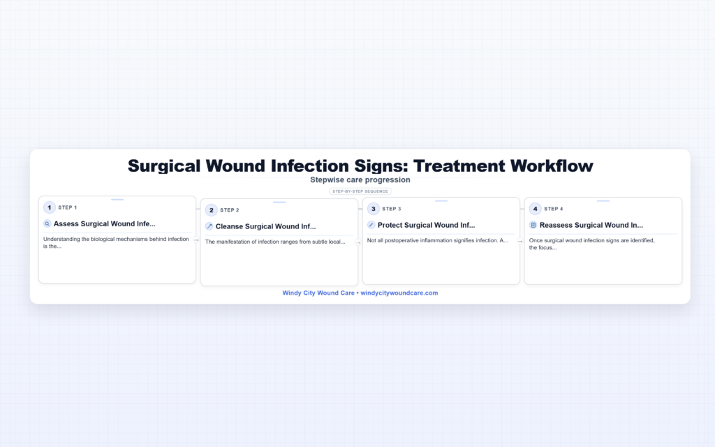
Diagnostic Evaluation and Management
Once surgical wound infection signs are identified, the focus shifts to confirming the diagnosis and determining the extent of the infection. A systematic approach ensures that appropriate cultures are obtained before the administration of antibiotics, allowing for targeted therapy. Management often requires a combination of surgical intervention and pharmacological treatment tailored to the specific pathogens involved.
Laboratory and Microbiological Analysis
Laboratory tests are adjuncts to clinical judgment. Leukocytosis with a left shift is common in bacterial infections, but normal white blood cell counts do not rule out an SSI, especially in chronically ill patients. Inflammatory markers such as C-Reactive Protein (CRP) and Erythrocyte Sedimentation Rate (ESR) are elevated postoperatively but should trend downward; a secondary rise often heralds infection. The gold standard for diagnosis remains culture of wound aspirate or tissue biopsy. Superficial swabs are often contaminated by skin flora and are generally discouraged; deep tissue cultures provide more actionable data. According to the CDC, standardized surveillance definitions are essential for accurate reporting and treatment.
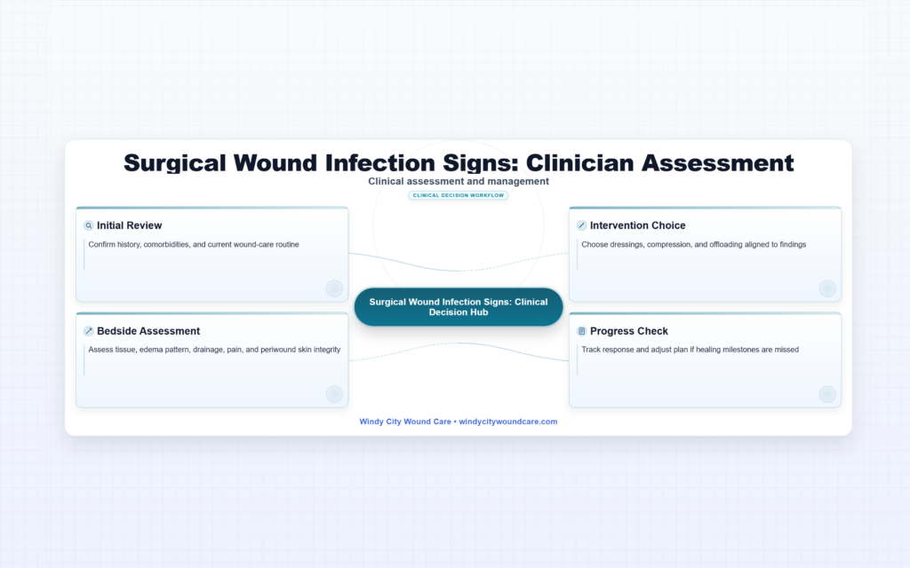
Clinical Evaluation Protocol
Assessment should be methodical to ensure no detail is overlooked. The following protocol outlines the steps for evaluating a suspected SSI:
- History Review: Document the timing of symptom onset, note any comorbidities like diabetes, and review the operative note for potential contamination levels.
- Physical Examination: Remove dressings using aseptic technique. Measure the area of erythema. Assess for fluctuance or crepitus (gas in tissues).
- Wound Exploration: If dehiscence is present or fluctuance is noted, probe the wound with a sterile blunt instrument to check for fascial involvement.
- Specimen Collection: Obtain deep tissue cultures or fluid aspiration before initiating antibiotics.
- Imaging: If deep abscess or necrotizing fasciitis is suspected, order a CT scan or ultrasound to delineate fluid collections.
Management strategies vary based on the depth of infection. Superficial incisional SSIs may only require local wound care and oral antibiotics. In contrast, deep or organ space infections often necessitate surgical debridement, opening of the wound, and parenteral antibiotics. For more detailed procedures on wound care, see our [internal link]wound debridement protocols[/internal link]. Additional resources can be found at the Mayo Clinic or NIH.
Frequently Asked Questions
What are the first signs of surgical wound infection?
Initial signs typically include increasing redness spreading from the incision, swelling, warmth, and pain that worsens rather than improves over time.
Is redness around a surgical incision normal?
Mild redness immediately adjacent to the suture line is normal during the inflammatory healing phase. However, expanding redness, streaking, or significant heat indicates infection.
How long after surgery can a wound infection occur?
Most infections occur within 30 days, but infections involving implants or prosthetics can manifest months or even a year after the procedure.
When should antibiotics be started for a wound infection?
Antibiotics should be initiated after cultures are obtained if there is clinical evidence of systemic infection, cellulitis, or purulent discharge. Superficial contamination may be managed with local care alone.
References & Resources
- CDC Surgical Site Infection Guidelines
- Mayo Clinic – Surgical Site Infection
- NIH – Wound Healing and Management